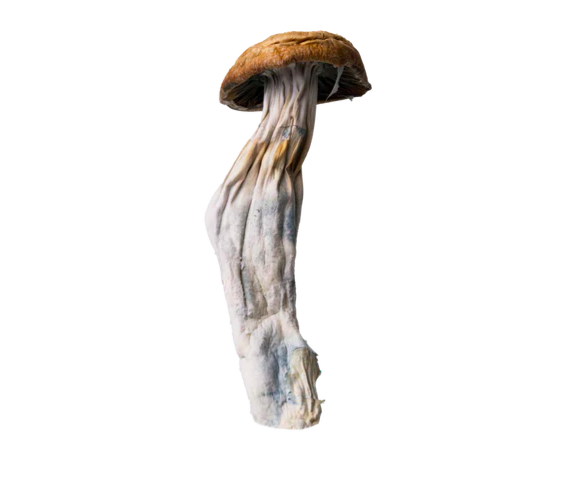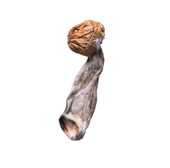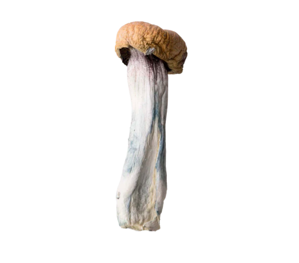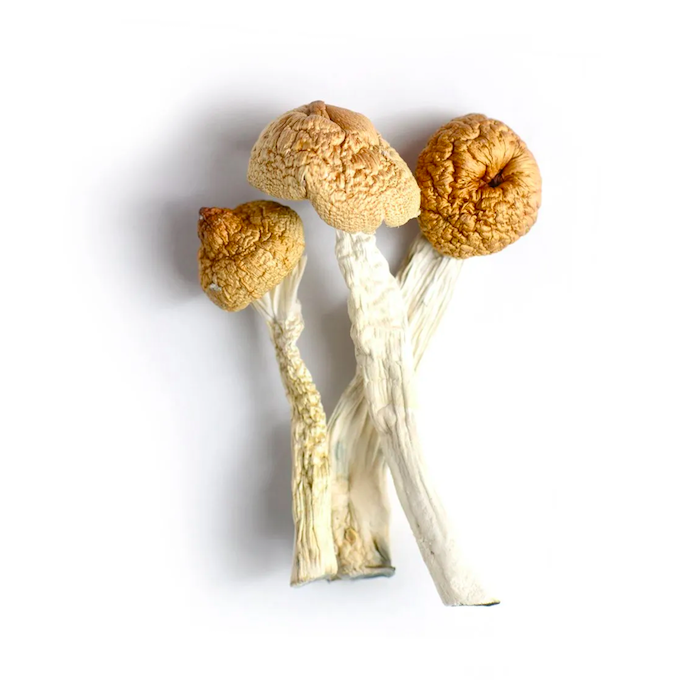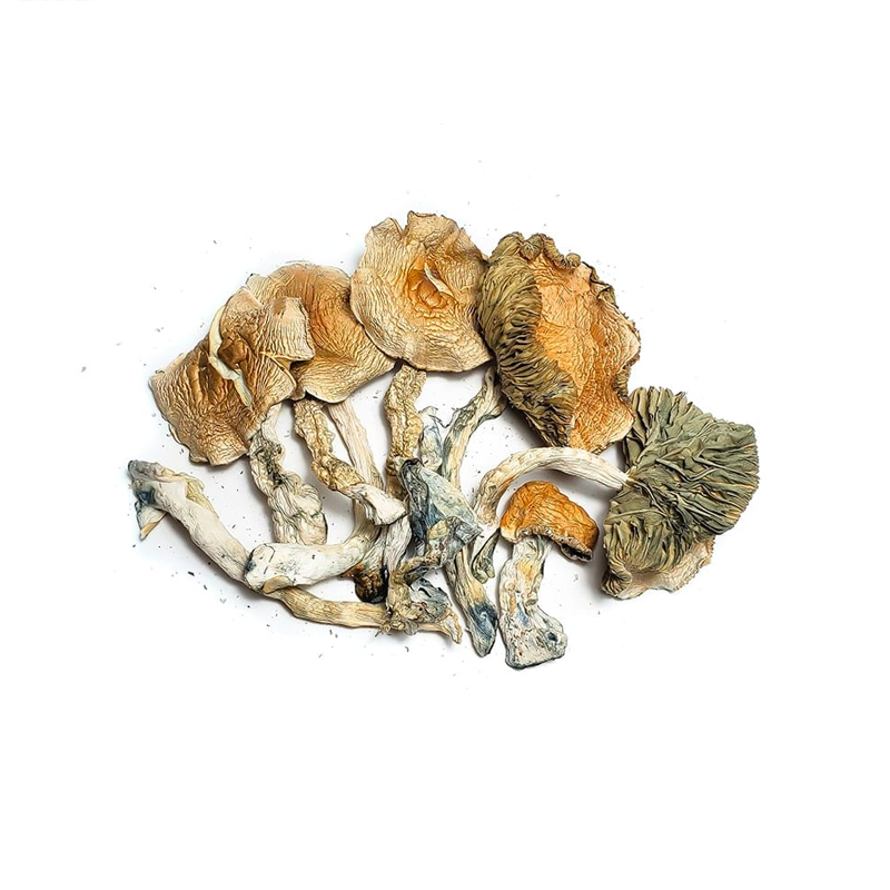
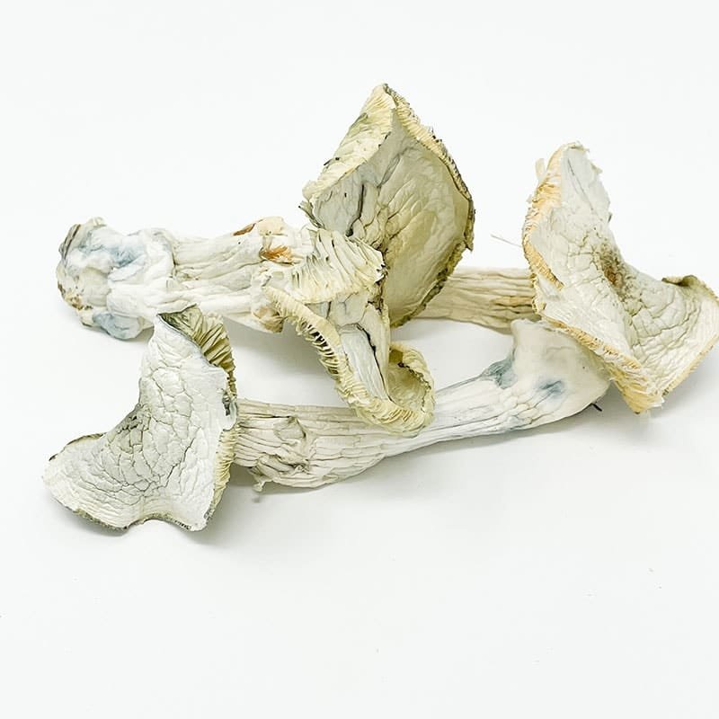
Albino A Mushrooms: Leucism, Genetics, and Mycological ResearchAlbino A mushrooms are a leucistic strain of Psilocybe cubensis characterized by reduced pigmentation in the fruiting body while retaining dark purple-brown spores. Studied for genetic variation, pigmentation pathways, and microscopic spore morphology, Albino A mushrooms are valuable reference specimens in fungal taxonomy, developmental biology, and comparative mycology research.What Are Albino A Mushrooms?Albino A mushrooms are a leucistic strain of Psilocybe cubensis defined by suppressed pigmentation in somatic tissues — pileus and stipe — alongside fully pigmented dark purple-brown basidiospores. This phenotypic asymmetry between pale fruiting body and dark reproductive spores is the defining biological signature of the AA mushroom strain and the primary reason it holds sustained scientific value in fungal genetics, taxonomy, and microscopy research.Core definition: Albino A mushrooms are not true albinos. They are leucistic — meaning pigment-producing capacity is reduced in specific tissues, not eliminated system-wide. This single distinction separates Albino A from genuinely achromatic fungal variants and determines every downstream conclusion about their genetics, classification, and research application.Key Facts: Albino A MushroomsAttributeValueCommon NameAlbino A (AA )SpeciesPsilocybe cubensisFamilyHymenogastraceaeOrderAgaricalesPigmentation TypeLeucistic (tissue-specific suppression)Spore ColorDark purple-brownSpore ShapeEllipsoid to subellipsoidApproximate Spore Size11–17 × 7–10 μmBruising ResponseBlue-green (indole oxidation)Phenotypic StabilityHigh — consistent across generationsPrimary Research ValuePigmentation pathways, taxonomy, microscopy, geneticsKey Distinguishing FeaturePale fruiting body with fully pigmented sporesClassification StatusIntraspecific strain of P. cubensisAlbino A mushrooms are a leucistic variant of Psilocybe cubensis examined across fungal taxonomy, developmental genetics, and microscopy research. Unlike true albino organisms — which lack all functional pigment-producing enzyme activity — Albino A mushrooms retain the biological capacity to synthesize melanin in their basidiospores despite producing visibly pale fruiting bodies. This tissue-specific pigmentation asymmetry is the strain’s defining scientific characteristic: it isolates melanin suppression to somatic developmental stages while preserving it in reproductive structures, providing researchers with a naturally occurring model for studying tyrosinase-dependent pigmentation regulation in Basidiomycota. The strain’s phenotypic stability across successive culture generations further supports its use as a reproducible laboratory reference. Mycologists studying Albino A mushrooms gain direct observational access to the boundary between leucistic suppression and functional melanin synthesis — a boundary with implications for fungal developmental biology, intraspecific taxonomy, and the comparative analysis of pigmentation genetics within Psilocybe cubensis.Scientific ClassificationRankClassificationKingdomFungiPhylumBasidiomycotaClassAgaricomycetesOrderAgaricalesFamilyHymenogastraceaeGenusPsilocybeSpeciesPsilocybe cubensisStrainAlbino A (AA )Pigmentation CategoryLeucistic variantTaxonomic StatusIntraspecific strain designationOriginal Species DescriptionFranklin Sumner Earle, 1906Reclassification BasisMolecular phylogenetic analysisIntroductionNot all mycological significance derives from geographic rarity or newly discovered taxa. Albino A mushrooms demonstrate that phenotypic variation within a well-documented species can generate research questions as substantive — and as precisely answerable — as those posed by novel species descriptions.Albino A mushrooms are a leucistic mutation of Psilocybe cubensis in which pigmentation is suppressed in the fruiting body while remaining fully functional in basidiospore production. This asymmetry positions the strain as a natural experimental model for one of the more tractable questions in fungal developmental biology: how is melanin synthesis regulated across different tissue types within a single organism, and what genetic mechanisms enforce that differential?The scientific significance of Albino A genetics and Psilocybe cubensis Albino A pigmentation pathways extends well beyond visual novelty. The strain raises concrete, answerable questions: Which regulatory mechanisms suppress tyrosinase activity in the pileus while preserving it in the basidium? What does tissue-specific leucism reveal about the developmental architecture of melanin biosynthesis in Basidiomycota? How does Albino A compare phenotypically and genetically to near-albino strains like Avery’s Albino and standard pigmented strains like Golden Teacher?This article addresses those questions systematically — through taxonomy, morphology, pigmentation biology, microscopy methodology, growth parameters, comparative strain analysis, and identification protocol — to establish a rigorous, citation-ready scientific reference for mycologists, researchers, and advanced students approaching Albino A mushrooms as subjects of formal inquiry.Understanding Leucism in Albino A MushroomsWhat Is Leucism in Fungi?Definition: Leucism is a genetic condition producing reduced pigment output in specific fungal tissues without disabling the organism’s melanin-synthesizing capacity system-wide.Key distinctions:Leucistic fungi retain functional tyrosinase enzyme activity in reproductive structuresPigmentation suppression is tissue-specific, not organism-wideSpores in leucistic strains retain normal dark colorationBruising responses remain intact, confirming separate biochemical pathwaysScientific implication: Leucism in fungi is a developmental regulatory phenomenon, not an enzymatic failure — making it a productive model for gene expression research rather than simply a pigmentation anomaly.This definition carries a critical taxonomic consequence: leucistic organisms are not albino. True albinism involves complete loss of functional tyrosinase — the enzyme catalyzing melanin synthesis — resulting in total pigment absence across all tissues and reproductive structures. In Albino A mushrooms, tyrosinase remains active in spore-forming cells, producing the fully pigmented basidiospores that confirm leucistic rather than albino status. A researcher who conflates these two conditions will arrive at taxonomically unsound conclusions about genetic mechanism, enzymatic function, and strain classification.Tyrosinase and Fungal Pigmentation PathwaysFungal pigmentation in Psilocybe cubensis is primarily regulated through tyrosinase-dependent enzymatic pathways. Tyrosinase catalyzes the oxidation of phenolic compounds — specifically L-DOPA and related precursors — into melanin intermediates that deposit as dark pigment in fungal tissue. This pathway operates across multiple tissue compartments during fruiting body development, with expression levels varying by tissue type, developmental stage, and genetic regulatory context.In Albino A mushrooms, current evidence indicates that tyrosinase activity is downregulated or transcriptionally suppressed in pileus and stipe tissues during fruiting body maturation, while remaining fully active in the basidium during basidiospore formation. This differential — somatic suppression alongside reproductive preservation — constitutes the mechanistic definition of the Albino A leucistic mutation and the primary justification for its use in Albino A fungal pigmentation research.Three candidate mechanisms may account for this tissue-specific suppression:Tissue-specific promoter activity regulating tyrosinase gene transcription in somatic versus reproductive compartmentsDifferential substrate availability — reduced melanin precursor supply in pileus tissue during specific developmental windowsPost-translational modification of tyrosinase enzyme activity varying by tissue type and developmental stageResolving which mechanism operates — or in what combination — requires targeted transcriptomic analysis of Albino A specimens at multiple developmental stages, representing a tractable and scientifically productive research program.Morphological Characteristics of Albino A MushroomsPileus (Cap)Mature Albino A mushrooms produce caps ranging from bright white to pale cream or ivory — a direct macroscopic expression of tyrosinase suppression in pileus tissue. This stands in contrast to the caramel-to-golden-brown coloration of standard Psilocybe cubensis strains, making cap color the first and most immediately recognizable identification marker for the strain.Key pileus characteristics:Color range: White to pale cream; ivory in some environmental conditionsSurface texture: Smooth under high humidity; may develop fine surface cracking under low-humidity conditionsDevelopmental profile: Convex to broadly umbonate in young specimens; progressively flattening with maturityBruising: Blue-green discoloration at mechanical damage sites — confirming indole compound oxidation is biochemically independent of melanin suppressionMargin: Slight inward curl in early development; expanding and flattening with spore maturityThe persistence of blue-green bruising in Albino A pileus tissue is taxonomically and biochemically significant: it confirms that the indole oxidation pathway responsible for bruising operates independently of the tyrosinase-melanin pathway responsible for pigmentation, validating leucism as a pigmentation-specific modification rather than a broad enzymatic impairment.Stipe (Stem)The stipe of Albino A mushrooms presents as consistently pale — white to off-white — with a fibrous longitudinal texture. An annulus derived from the partial veil is present in younger specimens, degrading as cap expansion progresses. Stipe dimensions fall within ranges typical for Psilocybe cubensis, with substrate richness, temperature, and humidity influencing final length and diameter. Bruising behavior consistent with pileus response is present on stipe tissue when mechanically disturbed.Lamellae (Gills)Gill coloration in Albino A mushrooms progresses from pale gray in immature specimens to deep purple-brown at full basidiospore maturity. This darkening — driven by spore deposition on gill surfaces — occurs across Albino A specimens despite their pale somatic phenotype, providing direct visual confirmation that spore pigmentation operates through a regulatory pathway unaffected by the leucistic mutation. Gills are closely spaced and adnate to adnexed in stipe attachment, consistent with Psilocybe cubensis species morphology.Spore PrintThe spore print of Albino A mushrooms is deep purple-brown to near-black — fully consistent with Psilocybe cubensis at the species level. A dark spore print from a white-capped specimen is the single most diagnostically significant macroscopic observation in Albino A identification: it confirms leucistic rather than albino status, distinguishes the strain from genuinely achromatic variants, and anchors its classification as an intraspecific pigmentation variant rather than a taxonomically separate entity.Quick Identification Checklist: Albino A MushroomsFor field and laboratory identification, apply this five-point diagnostic sequence:☐ Cap color: White to pale cream — absent golden-brown pigmentation of standard P. cubensis strains☐ Bruising response: Blue-green discoloration at mechanical damage sites on cap and stem — confirms species-level identity☐ Gill darkening: Progressive purple-brown coloration as specimens mature — pale body with darkening gills is strongly diagnostic☐ Spore print: Deep purple-brown to near-black — definitive confirmation of leucistic over true albino status☐ Microscopic verification: Ellipsoid spores, thick smooth walls, visible apical germ pore — confirms P. cubensis species identityDecision rule: All five markers present = Albino A leucistic identification confirmed. Pale spore print despite pale body = consider Avery’s Albino or true near-albino variant.Albino A Spore Morphology and Microscopy StudyBasidiospore Reference DataCharacteristicDescriptionShapeEllipsoid to subellipsoidWallThick, smooth, without surface ornamentationColor in massDark purple-brownColor individuallyGolden to amber under transmitted lightGerm poreProminent, apical, clearly visible at ×400–×1000Approximate length11–17 μmApproximate width7–10 μmLength-to-width ratioApproximately 1.5–1.7:1SymmetrySlightly inequilateral in lateral viewThe full pigmentation of Albino A basidiospores under microscopy — dark purple-brown in mass, amber individually under transmitted light — stands in direct, measurable contrast to the pale macroscopic phenotype of the fruiting body. This contrast is the central biological phenomenon driving Albino A microscopy study: researchers can directly observe and document the developmental point at which pigmentation suppression ends and melanin synthesis resumes in spore-forming tissue.Microscopy Workflow: Step-by-Step ProtocolObjective: Characterize Albino A spore morphology and gill anatomy under light microscopy for taxonomic documentation and comparative research.Equipment required:Compound light microscope (minimum ×400 magnification; ×1000 oil immersion preferred)Glass slides and coverslips3% KOH mounting medium or water mountSpore print or fresh gill sectionStage micrometer for spore measurement calibrationBrightfield and phase contrast illuminationProtocol:Step 1 — Spore preparation: Transfer a small quantity of spore print material to a glass slide using a sterile loop. Apply one drop of 3% KOH or distilled water. Lower coverslip at angle to eliminate air bubbles.Step 2 — Initial examination (×100–×200): Survey the slide for spore distribution, clumping patterns, and contamination. Confirm purple-brown mass coloration consistent with Albino A spore morphology.Step 3 — Morphological characterization (×400): Identify individual spores. Confirm ellipsoid to subellipsoid shape, smooth thick walls, and absence of surface ornamentation. Note coloration under transmitted light (golden to amber).Step 4 — Germ pore confirmation (×400–×1000): Focus to the apical spore terminus to identify and document the prominent germ pore. Its presence and structural integrity confirm species-level Psilocybe cubensis identification.Step 5 — Spore measurement: Using a calibrated stage micrometer, measure a minimum of 20 individual spores for length and width. Record mean, range, and length-to-width ratio for database entry and comparative reference.Step 6 — Gill section analysis: Prepare a thin hand-section of gill tissue. Mount in KOH. At ×400–×1000, identify basidia (club-shaped, four sterigmata), cheilocystidia (fusiform to lageniform), and spore attachment points. Document findings against species-level descriptions.Interpretation standard: Spore measurements falling within 11–17 × 7–10 μm, combined with prominent germ pore, smooth thick wall, and purple-brown mass coloration, confirm Psilocybe cubensis species identity in Albino A specimens.Basidia and CheilocystidiaGill sections from Albino A mushrooms reveal basidia as club-shaped structures bearing four sterigmata, each supporting a single basidiospore. Cheilocystidia — sterile cells lining gill edges — present fusiform to lageniform morphology consistent with Psilocybe species descriptions. These cellular structures are taxonomically continuous with standard P. cubensis anatomy, confirming that the leucistic mutation in Albino A is a pigmentation-specific modification that does not alter fundamental gill architecture or spore-bearing cell morphology.Albino A Growth ParametersUnderstanding Albino A growth parameters contextualizes the strain’s morphological expression within its developmental environment and provides the quantitative reference data necessary for comparative mycological study.Environmental and Developmental Reference TableParameterDocumented RangeNotesOptimal fruiting temperature22–26°C (72–79°F)Consistent with P. cubensis species rangeColonization temperature28–30°C (82–86°F)Mycelium growth phaseRelative humidity (fruiting)90–95%High humidity preserves pale cap surface integritySubstrate compatibilityBrown rice flour, rye grain, pasteurized straw, manure-basedStandard P. cubensis substrate rangeColonization speedModerate — comparable to other P. cubensis strainsNo documented significant deviation from species averageMycelium appearanceWhite to off-white; rhizomorphic growth pattern commonConsistent with leucistic pigmentation phenotypeFirst primordia formationTypically 1–3 weeks post-colonizationEnvironment-dependentPhenotypic stability across generationsHighLeucistic trait reliably expressed across culturesResearch note on colonization speed: Albino A colonization speed is frequently discussed in mycological communities as a distinguishing characteristic. Current observational data suggests colonization rate is broadly comparable to standard P. cubensis strains under equivalent environmental conditions — indicating that the leucistic mutation does not confer a measurable metabolic advantage or disadvantage during mycelium establishment. Formal controlled studies comparing Albino A colonization rate against standard strains under standardized substrate and temperature conditions would strengthen this conclusion.Albino A Genetics and Pigmentation ResearchGenetic Basis of Leucism in Albino A The Albino A leucistic mutation involves genetic modification of pigmentation regulatory pathways rather than deletion of melanin-synthesizing genes. Current understanding supports three candidate mechanisms operating at the molecular level:Transcriptional regulation: Tissue-specific promoter elements suppressing tyrosinase gene expression in somatic fruiting body tissues while maintaining transcriptional activity during sporogenesisSubstrate limitation: Reduced availability of melanin precursor compounds — L-DOPA and related phenolics — in pileus tissue relative to basidial cells during spore maturationPost-translational control: Enzymatic modification of tyrosinase protein activity that varies across tissue types, potentially through differential cofactor availability or inhibitory protein interactionThe fact that Albino A phenotypic stability is high across successive culture generations indicates genetic encoding at a stable locus rather than epigenetic or environmentally induced modification — a distinction that significantly increases the strain’s value as a controlled research model for Albino A genetics investigation.Phenotypic Stability as a Research AssetThe consistent expression of leucistic traits across Albino A mushroom generations — pale fruiting body, dark spores, intact bruising response — represents a measurable research asset. Phenotypically stable strains allow researchers to establish reliable baseline measurements, replicate observations across laboratory settings, and build comparative datasets without confounding variation from inconsistent phenotype expression. For these reasons, phenotypic stability in Albino A mushrooms is not merely a descriptive characteristic but a methodological advantage for mycological research programs.Comparative Analysis: Albino A vs. Avery’s Albino vs. Golden TeacherThree-Strain Comparison MatrixCharacteristicAlbino A Avery’s AlbinoGolden TeacherPigmentation typeLeucisticNear-albino to leucisticStandard pigmentedCap colorWhite to pale creamWhite to ghostly paleGolden-brown to amberSpore print colorDark purple-brownVery pale to near-colorlessDark purple-brownSpore pigmentationFully retainedSignificantly reducedFully retainedBruising responseBlue-green (present)Blue-green (present)Blue-green (present)Phenotypic stabilityHighVariable (reported)HighColonization speedModerateModerateModerate to fastMycelium colorWhite to off-whiteWhiteWhitePrimary research valueTissue-specific leucism modelNear-albino phenotype referenceStandard pigmented baselineTaxonomic statusP. cubensis strainP. cubensis strainP. cubensis strainKey differentiatorDark spores despite pale bodyReduced spore pigmentationFull pigmentation throughoutBest used forPigmentation pathway contrast studiesNear-albino genetic analysisComparative baselineAlbino A vs. Avery’s Albino: The Critical DistinctionThe fundamental differentiator between Albino A vs. Avery’s Albino is spore pigmentation. Albino A produces fully pigmented dark purple-brown spores — confirming that melanin synthesis remains functional in reproductive structures — while Avery’s Albino exhibits dramatically reduced spore coloration approaching near-achromia. This difference makes Albino A the more experimentally tractable model for tissue-specific leucism research: the contrast between pale somatic tissue and dark spores is unambiguous, measurable, and reproducible. Avery’s Albino, with its more generalized pigmentation reduction, presents a less clearly defined phenotypic boundary for comparative analysis.Albino A vs. Golden Teacher: Establishing the BaselineGolden Teacher represents the standard pigmented Psilocybe cubensis reference against which leucistic strains are most productively compared. Both strains produce dark purple-brown spore prints and exhibit identical bruising responses — confirming shared species-level biochemistry. The critical difference is macroscopic cap pigmentation: Golden Teacher expresses full tyrosinase-dependent melanin deposition across somatic tissues, while Albino A suppresses this deposition in the pileus and stipe. Paired laboratory observation of Albino A and Golden Teacher specimens provides the most direct visualization of leucism as a pigmentation-specific developmental modification within Psilocybe cubensis.Albino A Identification GuideMacroscopic Identification ProtocolReliable Albino A mushroom identification requires systematic multi-feature assessment. No single macroscopic characteristic — including cap color — is sufficient for confident strain confirmation.Step 1 — Pileus assessment: Confirm white to pale cream cap coloration without golden-brown pigmentation typical of standard P. cubensis. Document developmental stage and cap profile.Step 2 — Bruising verification: Apply gentle mechanical pressure to cap and stem tissue. Confirm blue-green bruising response. Absence of bruising suggests non-Psilocybe identification; presence confirms species-level candidate.Step 3 — Gill color progression: Observe lamellae color. Purple-brown darkening in a pale-bodied specimen is strongly diagnostic for Albino A leucistic phenotype. Pale or white gills without darkening raise consideration of alternative identification.Step 4 — Spore print collection: Rest mature cap gill-side down on paper in a still environment for 4–8 hours. A deep purple-brown to near-black print from a pale-capped specimen is the definitive macroscopic confirmation of leucistic over true albino classification.Step 5 — Microscopic verification: Mount spore material for light microscopy. Confirm ellipsoid to subellipsoid morphology, thick smooth walls, and visible apical germ pore at ×400–×1000 magnification. Measure a minimum of 20 spores for dimensional confirmation against the 11–17 × 7–10 μm reference range.Differentiating Albino A From Similar StrainsObservationSuggests Albino A Suggests Avery’s AlbinoSuggests Standard StrainCap colorWhite to pale creamWhite to ghostly paleGolden-brownSpore printDark purple-brownNear-colorless to very paleDark purple-brownGill darkeningProgressive, clearMinimal or reducedProgressive, clearBruisingPresentPresentPresentMicroscopic spore colorDark in mass, amber individuallyPale amber to near-clearDark in mass, amber individuallyAlbino A Mushrooms in Mycology ResearchContribution to Fungal TaxonomyAlbino A mushrooms reinforce a foundational principle of rigorous mycological taxonomy: pigmentation characteristics must be evaluated across multiple tissue types — somatic and reproductive — rather than assessed from macroscopic cap appearance alone. A researcher classifying Psilocybe cubensis specimens by cap color exclusively would systematically mischaracterize Albino A as a distinct taxonomic entity rather than an intraspecific leucistic variant. The strain therefore functions as a practical teaching case for taxonomic methodology as much as a research subject in its own right.Contribution to Developmental BiologyAlbino A mushrooms provide a naturally occurring model system for investigating tissue-specific gene expression in fungal development. The spatial restriction of pigmentation suppression — operating in somatic tissues while absent from spore-forming cells — implies that melanin biosynthesis in Psilocybe cubensis is governed by developmental regulatory mechanisms that partition gene expression across tissue compartments. Characterizing these mechanisms through comparative transcriptomic analysis of Albino A specimens at multiple developmental stages would contribute directly to understanding how eukaryotic fungi coordinate gene expression across structurally and functionally distinct tissue types.Contribution to Fungal Biodiversity ResearchStable, morphologically characterized intraspecific variants like Albino A mushrooms provide anchoring reference data for fungal biodiversity databases and population genetics studies. As molecular tools make strain-level genetic characterization increasingly accessible, strains with well-documented and reproducible phenotypic signatures become integration points between morphological observation records and genomic datasets — strengthening the empirical infrastructure of Psilocybe cubensis biodiversity research and the broader field of Basidiomycota population genetics.FAQ: Albino A MushroomsWhat are Albino A mushrooms?Albino A mushrooms are a leucistic strain of Psilocybe cubensis defined by genetically suppressed pigmentation in somatic fruiting body tissues — producing white to pale cream caps and stems — alongside fully retained dark purple-brown basidiospores. This tissue-specific pigmentation asymmetry is the strain’s defining characteristic and primary scientific value: it isolates melanin suppression to specific developmental stages while preserving reproductive pigmentation, providing a tractable natural model for studying tyrosinase-dependent melanin regulation in Basidiomycota.Is Albino A a true albino strain?No — and the distinction is scientifically critical. Albino A is leucistic, not albino. True fungal albinism involves complete loss of functional tyrosinase enzyme activity, producing unpigmented spores alongside a pale fruiting body. Albino A retains full tyrosinase function in basidial cells during sporogenesis, generating dark purple-brown spores that confirm pigmentation capacity is intact. Misclassifying the strain as truly albino leads to incorrect conclusions about its genetics, enzymatic function, and taxonomic relationship to other pale-phenotype P. cubensis strains.Why are Albino A mushrooms white?The pale appearance of Albino A mushrooms results from genetically encoded suppression of tyrosinase-dependent melanin synthesis in pileus and stipe tissues during fruiting body development. This suppression is tissue-specific: it operates in somatic structures while leaving melanin production intact in spore-forming basidia. The mechanism is regulatory rather than enzymatic — tyrosinase is not absent in Albino A somatic tissue but is transcriptionally suppressed or post-translationally inhibited during the developmental window in which cap pigmentation would otherwise form.What color are Albino A spores?Albino A spores are dark purple-brown in mass — fully consistent with species-level Psilocybe cubensis characteristics. Individually, under transmitted light microscopy, they appear golden to amber. Full spore pigmentation in a pale-bodied strain is the definitive diagnostic marker confirming leucistic classification over true albinism, and represents direct evidence that melanin biosynthesis remains functional in Albino A reproductive structures.How are Albino A mushrooms studied scientifically?Albino A mushrooms are examined through: (1) light microscopy for basidiospore morphology, wall structure, germ pore characterization, and spore measurement; (2) gill section preparation for basidia and cheilocystidia analysis; (3) spore print assessment for pigmentation confirmation; (4) comparative morphological analysis against standard and near-albino P. cubensis strains; and (5) genetic and biochemical investigation of tyrosinase-related pigmentation regulatory pathways. Phenotypic stability across culture generations makes the strain a reproducible and methodologically reliable laboratory subject.What makes Albino A useful in mycology?Albino A mushrooms are scientifically useful because they provide a stable, naturally occurring model in which pigmentation suppression is confined to somatic tissues — enabling direct observational and experimental access to the boundary between leucistic suppression and functional melanin synthesis. Their consistent phenotypic expression supports reproducible laboratory study, their taxonomic continuity with P. cubensis provides a controlled comparison baseline, and their contrasting macroscopic and microscopic pigmentation patterns generate testable hypotheses in developmental genetics, comparative taxonomy, and fungal biodiversity research.What is leucism in fungi?Leucism in fungi is a genetic condition producing reduced or absent pigment output in fruiting body somatic tissues without disabling the organism’s melanin-synthesizing capacity in reproductive structures. Leucistic fungi retain functional tyrosinase activity in spore-forming cells, producing normally pigmented basidiospores despite a pale fruiting body phenotype. This distinguishes leucism from true albinism — which involves complete enzymatic failure of melanin synthesis across all tissue types — and establishes leucism as a tissue-specific developmental regulatory phenomenon rather than a system-wide biochemical impairment.What family does Albino A belong to?Albino A belongs to the family Hymenogastraceae within the genus Psilocybe, order Agaricales — a classification established through molecular phylogenetic analysis that repositioned Psilocybe cubensis from the previously assigned family Strophariaceae. The reclassification was driven by comparative DNA sequence analysis across Agaricales lineages and reflects the phylogenetic primacy of molecular data over earlier morphology-based classification systems in contemporary fungal taxonomy.ConclusionAlbino A mushrooms are a scientifically precise demonstration that phenotypic variation within a single fungal species can illuminate fundamental questions in developmental biology, genetics, and taxonomy. Their leucistic mutation — suppressing melanin synthesis in somatic tissues while preserving it in reproductive spores — is not a cosmetic anomaly but a genetically encoded regulatory phenomenon with measurable implications for how Psilocybe cubensis partitions gene expression across developmental stages.Three conclusions follow directly from the Albino A evidence base. First, pigmentation in Psilocybe cubensis is not a uniform species-wide characteristic but a developmentally regulated process subject to tissue-specific genetic control — a conclusion made visible in every pale fruiting body producing a dark spore print. Second, taxonomic classification of pale-phenotype fungal strains requires multi-feature assessment anchored in spore morphology and microscopic anatomy; macroscopic cap color alone is an insufficient and misleading classification criterion. Third, stable, well-characterized intraspecific variants like Albino A mushrooms carry scientific value proportional to the rigor with which they are documented — they become anchoring reference points for integrating morphological, ecological, and genomic datasets as molecular tools advance.The most precise description of Albino A mushrooms is also their most scientifically productive framing: a strain defined by what it withholds — melanin from the fruiting body — and what it preserves with complete fidelity: the biological capacity for pigment synthesis, expressed precisely where reproductive success requires it.